Call: +919826033365 07971891384( PIN:867)
 Send Inquiry
 Send Inquiry
Mi Series Radiographic X-Ray System
Price 750000 INR/ Piece
MOQ : 1 Piece
Mi Series Radiographic X-Ray System Specification
- Material
- Stainless steel and coated metal
- Condition
- New
- Technology
- Digital Radiography (DR)
- Portable
- Yes, mobile trolley enabled
- Light Source
- High Intensity LED Collimator
- Pressure Range
- Atmospheric (for normal operations)
- Operating Temperature
- 5C - 40C
- Humidity %
- 80% RH (non-condensing)
- Sensors Specification
- Digital Flat Panel Detector, High Sensitivity, Large Active Area
- Indicator Specification
- LED status indicators for power and exposure
- Darkness Range
- Not Applicable (Digital output)
- Speed Range
- Auto imaging with rapid exposure cycle
- Ray Frequency Range
- 40-120 kHz X-ray generation
- Exposure Time Range (in Sec)
- 0.01 - 3 seconds adjustable
- Focus
- Adjustable focal spot, typically 1.0/2.0 mm
- Image System
- Digital image acquisition and storage
- Usage
- Hospital
- Power Source
- Electric
- Power Consumption
- 4 kW
- Working Voltage
- 220V AC, 50/60 Hz
- Dimension (L*W*H)
- Approx. 1800 x 700 x 1700 mm
- Weight
- Approx. 100 kg (varies with configuration)
- Image Storage Capacity
- 100,000 images (DICOM),
- Safety Features
- Automatic exposure control, overheat protection
- Max Output Power
- 5 kW
- Tube Current
- 10-200 mA (adjustable)
- Control Type
- Microprocessor based console
- User Interface
- Touch screen digital panel
- Image Display
- 17" high-resolution medical monitor
- Tube Voltage
- 40-125 kV (adjustable)
- Patient Table
- Height adjustable, radiolucent design
- Networking
- PACS compatible, Ethernet/Wi-Fi connectivity
Mi Series Radiographic X-Ray System Trade Information
- Minimum Order Quantity
- 1 Piece
- Payment Terms
- Cash Advance (CA)
- Supply Ability
- 5000 Pieces Per Week
- Delivery Time
- 2-7 Days
- Main Domestic Market
- All India
About Mi Series Radiographic X-Ray System
Empowered with a team of passionate personnel, we are engaged in offering Mi Series Radiographic X-Ray System. Our offered system is used to diagnose or treat patients by recording images of the internal structure of the body. This Mi Series Radiographic X-Ray System is made available in different specifications for our clients to choose from.
Advanced Digital Imaging Technology
Utilizing state-of-the-art Digital Radiography (DR), the Mi Series ensures sharp, high-contrast images with rapid exposure and auto imaging features. The digital flat panel detector, combined with high output power and adjustable focus, delivers consistent results in varying clinical scenarios.
Intuitive User Experience
A microprocessor-based digital console powered by a touchscreen panel streamlines the imaging process. LED indicators, automatic exposure controls, and preset safety protocols simplify operation, letting clinicians focus on patient care and precision.
Comprehensive Connectivity & Storage
The system supports Ethernet and Wi-Fi connectivity, enabling seamless integration with hospital PACS. Store up to 100,000 DICOM-format images and view results instantly on a 17-inch medical-grade monitor for efficient case management.
FAQ's of Mi Series Radiographic X-Ray System:
Q: How does the Mi Series X-Ray System enhance workflow in hospitals?
A: The system streamlines workflow through its intuitive touch screen control panel, rapid auto imaging capabilities, and large image storage capacity. Combined with PACS compatibility and wireless connectivity, it offers quick access to images for diagnosis and sharing across departments.Q: What safety features are included in this digital radiography system?
A: Safety is ensured with built-in automatic exposure control, overheat protection, LED status indicators, and a radiolucent patient table, minimizing risks for both patients and operators during radiographic procedures.Q: When should the adjustable tube voltage and current be changed?
A: Adjust the tube voltage (40-125 kV) and current (10-200 mA) based on specific imaging requirements, patient size, or clinical protocols to optimize image clarity while managing radiation dose. The microprocessor console simplifies these adjustments for the operator.Q: Where can acquired images be viewed and stored within the system?
A: Captured images appear instantly on a 17-inch high-resolution medical monitor and are digitally stored onboard with capacity for 100,000 DICOM images. The system supports both local review and networked sharing via PACS through Ethernet or Wi-Fi.Q: What is the process for operating the Mi Series X-Ray System?
A: Operation involves positioning the patient on the height-adjustable, radiolucent table, setting exposure parameters via the touchscreen control, and initiating imaging. The automatic exposure control and rapid cycle speed ensure efficient and consistent acquisition.Q: How portable is the Mi Series X-Ray System?
A: Despite its robust stainless steel construction, the system is designed for mobility with a trolley-enabled chassis, making it suitable for bedside imaging and easy transportation within hospital settings.Q: What benefits does the high-intensity LED collimator provide?
A: The high-intensity LED collimator ensures precise beam alignment and field illumination, improving imaging accuracy, reducing retakes, and enhancing patient safety during each X-ray procedure.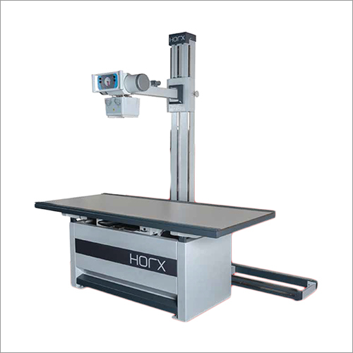
Tell us about your requirement

Price:
Quantity
Select Unit
- 50
- 100
- 200
- 250
- 500
- 1000+
Additional detail
Mobile number
Email

More Products in X-Ray System Category
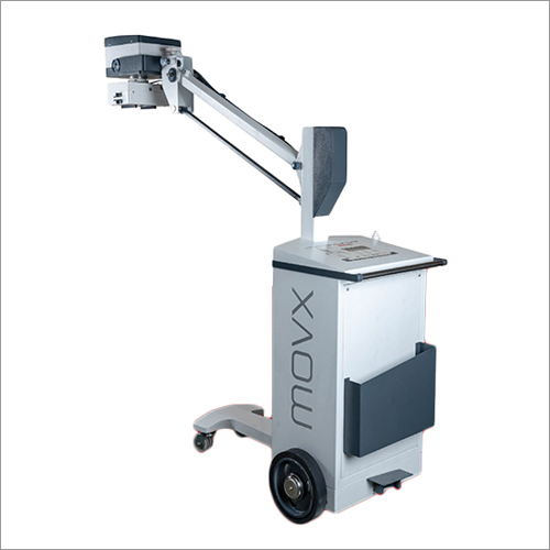
Mobile X-Ray System
Price 195000 INR / Piece
Minimum Order Quantity : 1 Piece
Technology : Other, Digital Radiography (DR)
Exposure Time Range (in Sec) : 0.016 seconds adjustable
Operating Temperature : 10C 40C
Light Source : Other, LED Collimator Light
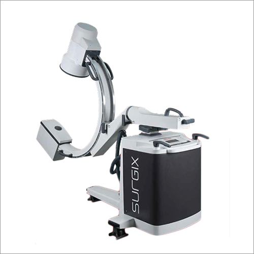
Mobile C-Arm Imaging System
Price 1100000 INR / Piece
Minimum Order Quantity : 1 Piece
Technology : Other, Digital Imaging, Fluoroscopy
Exposure Time Range (in Sec) : 0.16 sec
Operating Temperature : 5C to 40C
Light Source : Other, Highintensity Xray tube
Contact Details
DNHS HEALTHCARE DEVICES PRIVATE LIMITED
GST : 23AAHCD5042K1ZR
- Unit-77/1, Sanwer Road, Near Central Warehouse Narwal, Kumar Khadi,Indore - 452015, Madhya Pradesh, India
- Phone :+917971891384 PIN:( 867 )
Developed and Managed by Infocom Network Private Limited.
